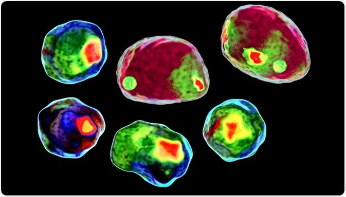
Flow cytometry

Optical filters are an essential component of flow cytometry, a technique used to measure and analyze the properties of individual cells or particles in a fluid stream. The primary function of optical filters in flow cytometry is to selectively transmit or block specific wavelengths of light, which helps to distinguish different cell populations based on their fluorescence characteristics.
Flow cytometry works by illuminating cells or particles with one or more lasers, and then detecting and measuring the light scattered or emitted by the cells. Optical filters are used in flow cytometry to selectively filter out unwanted wavelengths of light and allow only the relevant wavelengths to pass through to the detectors.
Flow cytometry uses several wavelengths of light to excite and detect fluorescent dyes or proteins in cells. The specific wavelengths used can vary depending on the type of dyes or proteins being analyzed.
For example, the most commonly used lasers in flow cytometry are 488 nm, 633 nm, and 405 nm, and they are used to detect commonly used dyes such as FITC, PE, APC, and Pacific Blue. However, some applications may require different or additional wavelengths, such as 561 nm or 594 nm for detecting red fluorescent proteins.
Therefore, the specified wavelength of flow cytometry depends on the specific experimental setup, the types of dyes or proteins used, and the desired detection sensitivity and resolution.
In addition to these standard wavelengths, flow cytometry can also use other wavelengths depending on the specific applications and types of dyes used.


